Pregnant women have to go through several ultrasounds for diagnoses and fetal development analysis. It’s the only chance for a woman to see their unborn child, hear reports on their health, and listen to their heartbeat. It can be a beautiful bonding moment for a mother-to-be.
Yet, healthcare specialists don’t often make more than two or three ultrasounds during pregnancy. So why is it so? Well, it appears that such a number is just what healthcare providers and expecting mothers need to receive all essential information about the developing baby. However, there are certain exceptions to this rule.
Overall, future mothers will most certainly take several ultrasounds during their pregnancies. So, it’s best to know how this diagnostic method works, how safe it is, and what to expect during these examinations.
How Does an Ultrasound Scan Work?
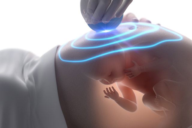
An ultrasound is a type of diagnostic imaging made with sound waves. In this method, high-frequency sound waves are used to create an image of the insides of a body. Of course, the sound itself can’t create images of your baby.
This technology relies on the echoes bouncing off organs to produce electrical signals. Further, the machine or computer interprets those signals, converting them into images. Next, an ultrasound technician will analyze the results and draw a conclusion based on the states of organs and tissues shown in the ultrasound.
An ultrasound is a non-invasive and safe method to receive valuable insights into one’s diagnosis or treatment.
However, it’s important to note that although ultrasound is considered completely safe, the medical community doesn’t have proof of that. So far, no signs of risks or side effects have been found. Yet, it doesn’t mean it will remain so in the future. So, what we know as of now, ultrasound is completely safe. Still, most healthcare specialists don’t turn to this diagnostic tool without real medical reasons.
The First Trimester Ultrasound
The first ultrasound is usually scheduled at 11-14 weeks of pregnancy. During this period, healthcare specialists can already see a fetus on the scan and can run the needed diagnostics. Still, sometimes women come for an early ultrasound at around 4-5 weeks. This is done to prove intrauterine pregnancy if a woman suspects pregnancy but her home pregnancy test or blood test leaves some uncertainty.
After positive results, women usually receive a referral for an ultrasound. Of course, at this point, an embryo has no heartbeat, nor is it visible on the ultrasound. Yet, a scan can show a gestational sac. This is a small structure development filled with amniotic fluid that normally occurs in a uterus during the first pregnancy weeks.
In more than 95% of cases, the discovery of a gestational sac speaks of a pregnancy. Such a structure is created to protect an embryo and provide it with nutrients via the yolk sac inside. Having more than one sac can signal two or more embryos. In addition, seeing the size of the gestational sac can help determine the conception date.
Unfortunately, in rare cases, ultrasound can’t find a gestational sac even if a woman has a positive pregnancy test. It usually means that a gestational sac traveled outside of the reproductive tract. This condition is called an ectopic pregnancy, and it requires immediate medical assistance. Such a pregnancy type will not result in birth.
However, healthcare specialists ask women for a second visit in one or two weeks to confirm the pregnancy. Thus, these women go through an additional ultrasound during their first trimester. The second scan ultrasound pictures should help doctors examine the fluids, placenta, and uterus, as well as fetus genetics and overall development.A doctor will check the following:
- pregnancy confirmation – vital signs check;
- normal development – a fertilized egg is attached to the uterus wall;
- determine a due date;
- detect the number of fetuses;
- rule over genetic abnormalities (i.e., a nuchal translucency screening).
The Second Trimester Ultrasound
Usually, a woman will have her second or third ultrasound already during her second trimester, around 18-20 weeks of pregnancy. This time difference leaves enough room for a fetus to develop. At this stage, the diagnostic imaging is called an anatomy scan. The name comes from the purpose of the scan. Typically, at the 20th week, an ob-gyn can determine the number of factors about the state of pregnancy, fetus development, sex, etc. A doctor will see:
- anatomic development of a fetus, including limbs, skull, spine, and inner organs;
- placenta position to determine the preferred delivery (i.e. a cesarean delivery);
- biological sex.
In general, during the second scan, doctors look for any potential birth defects, examine the vitals and healthy development of all organs and body parts of the baby, as well as monitor the mother’s health, and examine possible birth complications. One of them is a lowered placenta. It can result in difficult and dangerous birth. In such a case, more observations and scans will be recommended to track the situation and implement measures if needed.
Additional Ultrasounds
It’s worth noting that every woman and each pregnancy is different. So, it’s completely fine to attend ultrasound scans more than the assumed norm. Many factors, such as weight, age, bad habits (smoking, drinking), and personal or family history of certain health issues, may require additional monitoring.
Thus, women with obesity, diabetes, high blood pressure, or other diagnoses that put them into a high-risk group will have to go through an ultrasound more than two or three times during their pregnancy. Often, the frequency of such examinations grows closer to the due date.
This way, an ultrasound can confirm the good health of a woman and fetus. It will track fetal movement and assess amniotic fluid. An ob-gyn can also spot any health threats and call for an urgent intervention judging only by the data on the scan. Also, extra ultrasounds are required whenever a baby has any abnormalities in its development.
Concluding
An ultrasound is a standard and safe procedure for expecting mothers and their babies. Such diagnostic methods can help determine safe pregnancy and healthy fetus development. In addition, it’s a strong tool for discovering and monitoring any pregnancy complications and birth defects.
Normally, women don’t have to undergo an ultrasound more than two times during pregnancy. However, there is nothing wrong with having more referrals to ultrasound during any trimester. Such diagnostic processes don’t harm the baby but can enhance the chances of a successful pregnancy and easy birth.