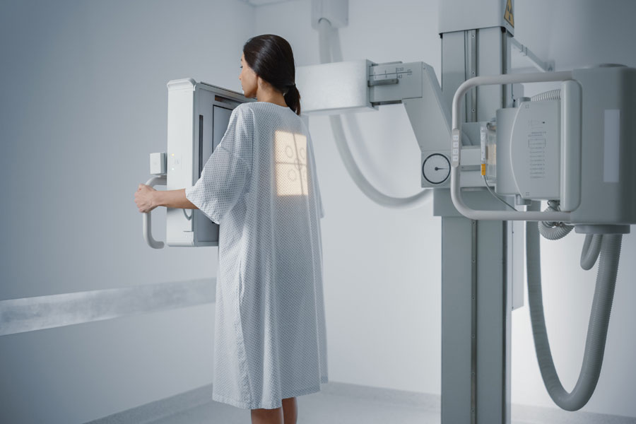
An x-ray examination is used to detect broken bones, abnormal tissue formations, such as cysts or tumors, lung lesions during pneumonia, injuries of a certain kind, the presence of foreign objects, dental problems, etc.
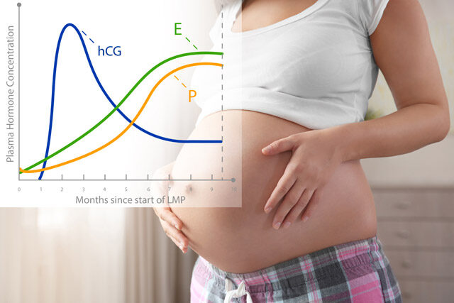
There is a common belief that x-ray screening is not safe during pregnancy which is not completely true. The American college of radiology states that the amount of radiation provided by a single screening cannot harm your unborn baby. However, guidelines regarding diagnostic imaging for pregnant women do exist.
How do x-rays work
X-rays are a type of electromagnetic radiation. When a patient gets a screening, they travel through their body, getting absorbed by different tissues in different amounts. This creates an image whether on a digital display or a piece of specially dedicated film depending on the type of the x-ray machine. This image provides an insight into what is happening inside the body.
Possible risks
The controversy about the increased risk of x-ray examination during pregnancy originates from the use of ionizing radiation. It is known to cause possible birth defects by affecting the fetus’s or embryo’s development in a negative way. This process is called teratogenesis. Radiation-induced teratogenesis may result in:
- loss of an embryo (on very early gestational period, before implantation);
- restricted growth or congenital anomalies in genitals, skeleton, or eyes (at 2–8 weeks after fertilization);
- microcephaly (too small head) and high to low risk of intellectual disabilities in the fetal period.
Does this sound scary? Yes, of course. But one should remember that this applies to very large doses of radiation: from 50 to 310 mGy and higher. In comparison, radiographic research of any extremity results in less than 0.001 mGy of fetal radiation dose. Even a pelvic CT scan which is a very specific use of ionizing radiation test performed close to the abdomen and can create a 50 mGy exposure, may be reduced to 2.5 mGy radiation by using a proper technique.
What you should do is discuss the situation both with your doctor and the radiologist so the scanning may be adjusted to your needs and carried out as safely as possible.
Speaking of the screening area, the mother-to-be and her child are absolutely safe if the reproductive system of the pregnant patient is protected from the ray beam. This means that the scans of the legs, head, teeth, or chest are not the slightest reason for concern. To be extra careful, your healthcare provider can give you a protective apron made of lead to ensure that you will not get abdominal x-rays. The same goes when you are needed to be present and help while your older child is getting an x-ray (e.g. to hold them) and there is nobody who can substitute you.
If you got screened not knowing about your pregnancy, you do not need to be alarmed: obstetricians and gynecologists agree that the radiation you get when diagnosed does not create an indication for abortion.
Alternative imaging techniques
The screening alternatives have their pros and cons. For example, an ultrasound, which is a routine part of regular pregnancy checkups, is safe for the baby even when performed on the abdominal area as it uses waves of sound. Another handy non-radioactive doctor’s tool is MRI which stands for magnetic resonance imaging. It can be more beneficial than ultrasound for deep screening and it does not depend on an operator.
But both of these methods are particularly effective for imaging soft or moving tissues and structures while x-rays are more suitable for scanning hard or air-filled parts of the body. Thus, when perfect to monitor a baby’s development, ultrasound is not an option for lung inflammation or possible bone fracture.
The takeaway
The ACOG (American College of Obstetricians and Gynecologists) confirms that, with rare exceptions, radiation exposure from a CT scan or an x-ray is of much lower dosage than amounts connected to teratogenesis.
Secondly, there can be situations when not having a procedure possesses more risk than the possible risk from radiation, and/or the respective equipment is more readily available. In the case of dangerous and acute processes, such as appendicitis, an early and precise diagnosis can be life-saving and, therefore, outweighs all the potential harm.
The key to having a safe x-ray without worrying about dangerous consequences is communication. Talk to your doctor, and discuss possible options so you will be able to develop a responsible and conscious solution that brings you maximum benefits with minimum risks. Remember that your health is of critical importance for the healthy development of your child so you should not neglect the required examinations when the situation calls for them.